本科留学生毕业设计说明书(论文)
Undergraduate International Students’ Graduation Project Report (Thesis)
题目: 基于多扫描仪数据的MRI快速重建方法研究
TITLE: RESEARCH ON TRAINING MRI RECONSTRUCTION NETWORK BASED ON MULTIPLE SCANNER DATA
Abstract
Paediatricians and paediatric endocrinologists utilize skeletal Bone Age Assessment (BAA) for investigations pertaining to genetic disorders, hormonal complications and abnormalities in the skeletal system maturity of children. Bone age conventional methods dating back to 1950 are often tedious and susceptible to inter-observer variability, and preceding attempts to improve these traditional techniques have inadequately addressed the human expert inter-observer variability so as to significantly refine bone age evaluations.
In this thesis, an automated and efficient approach with regression convolutional neural network is proposed. This approach automatically exploits the carpal bones as the region of interest (ROI) and performs boundary extraction of carpal bones, then based on the regression convolutional neural network it evaluates the skeletal age from the left hand wrist radiograph of young children from 0 to 6 years old. We also perform various experiments to determine whether it is more efficient to use the full hand radiographs or segmented regions of interest or the extracted carpal bones without tissue. Experiments show that the proposed method achieves an average discrepancy of 2.75 months between clinical and automatic bone age evaluations, and achieves 90.15% accuracy within 6 months from the ground truth for male, and 99.43% accuracy within 1 year.
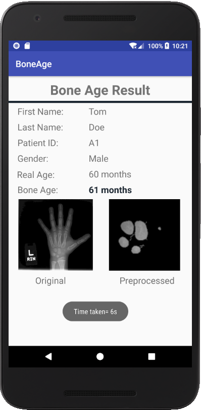
We then deploy this bone age software on a mobile application based on the Android operating system for personalized bone age assessments and a simplified Web Application for commercial entities. Our method poses a great potential for automated and more efficient and effective way of performing BAAs.
Key Words: bone age assessment, carpal bones segmentation, boundary extraction, regression convolutional neural network, mobile application
摘要
基于手骨X光片进行骨龄评估是儿科医生和儿科内分泌学专家诊断儿童发育疾病以及进行儿童生长发育评估常用的手段。传统的手骨骨龄评估方法,通常受限于医生的专业水平和自身经验,不同的医生得出的结论可能大不相同。尽管有不少方法尝试进行自动的骨龄评估,但依旧存在评估误差大的问题。
本文首先分析了手骨随着年龄增长的发育规律,发现不管男性还是女性,7岁前的掌骨骨块之间存在较大差异,具有明显的区分度,7岁后,掌骨骨块逐渐融合,区分度明显降低。因此,本文针对0~6岁儿童的生长发育,提出了一种自动有效的基于腕骨进行骨龄评估的方法。该方法利用腕骨作为感兴趣区,对腕骨进行边界提取,然后采用回归卷积神经网络对左手腕X线片进行年龄评估。同时为了验证腕骨在低龄儿童手骨骨龄预测中的有效性,本文进行了大量实验,对比了使用全手X光片,使用腕骨的分割区域,以及使用从腕骨区域提取的腕骨预测骨龄的结果。实验结果表明,本文提出的基于腕骨的评估方法性能最佳,该方法的骨龄评估结果与真实年龄之间的平均误差仅仅2.75个月,评估误差在6个月内的准确度达到90.15%,误差在1年内的准确度达到了99.43%。
然后,本文在以上算法研究的基础上,开发了基于Android操作系统的骨龄评估移动应用程序,用于个性化骨龄评估,并为商业实体开发了简化的Web应用程序。本文的方法和应用程序为自动有效地进行骨龄评估以及儿童生长发育评估提供了巨大的潜力。
关键词: X光片骨龄评估,腕骨分割,骨块边界提取,回归卷积神经网络,移动程序开发
Contents
1. Chapter I Introduction 3
1.1 Bone Age Assessments 3
1.2 Common Ways 3
1.3 Objective 5
1.4 The content of this thesis 5
2. Chapter II Related Works 6
2.1 Researches about CNN 6
2.1.1 Convolutional Neural Network Overview 6
2.1.2 CNN Methodology 6
2.2 Researches about BAA 9
3. Chapter III Proposed BAA Approach 12
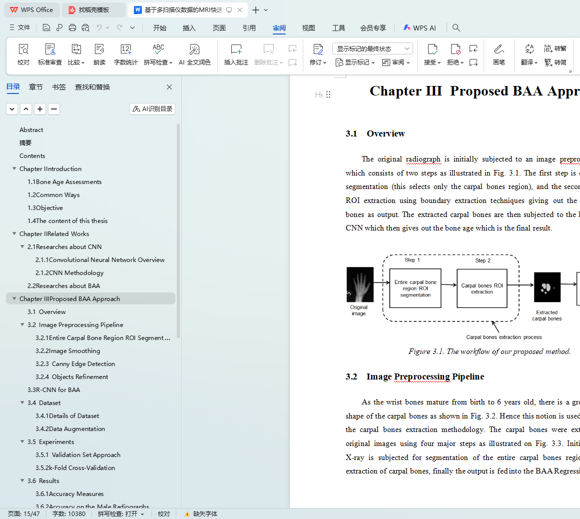
3.1 Overview 12
3.2 Image Preprocessing pipeline 12
3.2.1 Entire carpal bone region ROI segmentation 13
3.2.2 Image smoothing 14
3.2.3 Canny Edge Detection 14
3.2.4 Objects Refinement 14
3.3 R-CNN for BAA 15
3.4 Dataset 16
3.4.1 Details of Dataset 16
3.4.2 Data Augmentation 17
3.5 Experiments 17
3.5.1 Validation Set Approach 18
3.5.2 k-Fold Cross-Validation 18
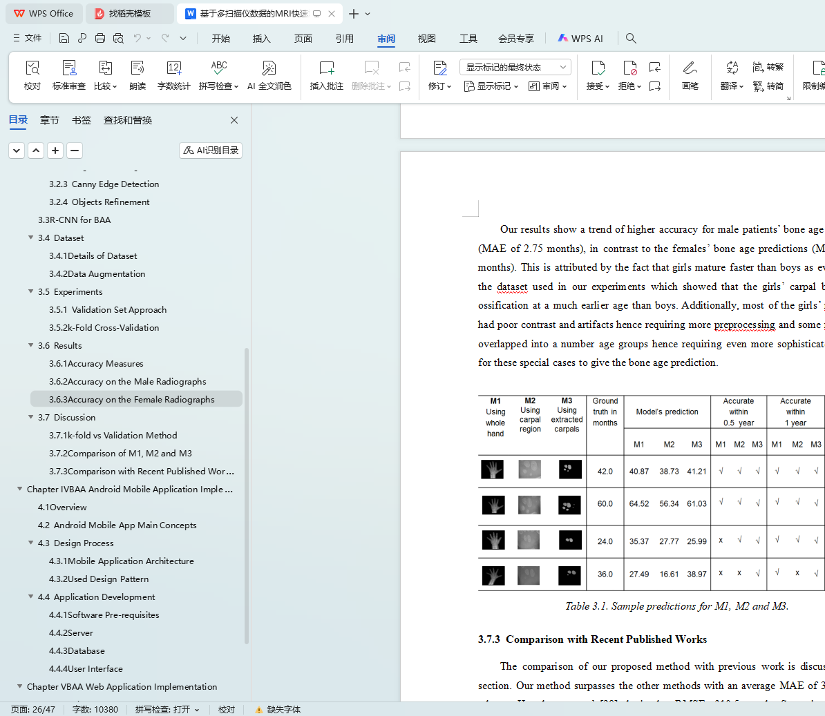
3.6 Results 19
3.6.1 Accuracy Measures 19
3.6.2 Accuracy on the Male Radiographs 19
3.6.3 Accuracy on the Female Radiographs 21
3.7 Discussions 22
3.7.1 k-fold vs Validation Method 22
3.7.2 Comparison of M1,M2 and M3 23
3.7.3 Comparison with recent published works 24
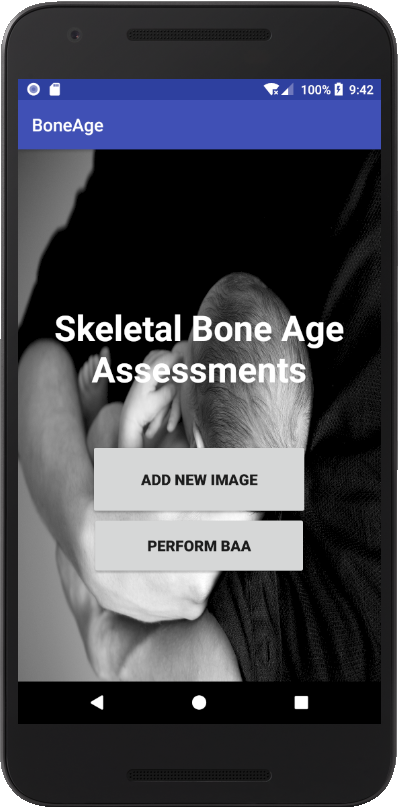
4. Chapter IV BAA Android Mobile App Implementation 27
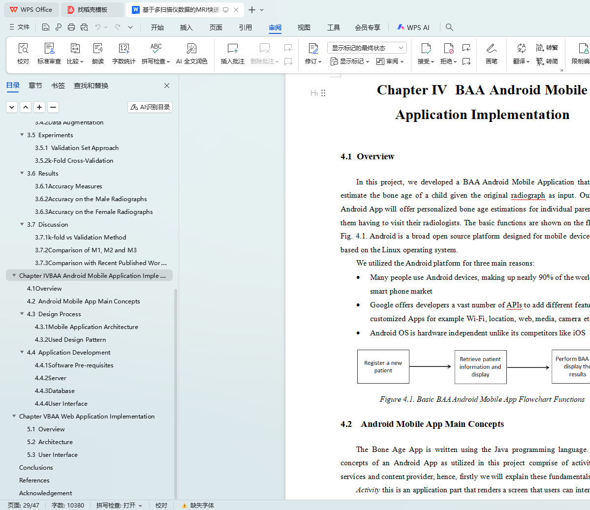
4.1 Overview 27
4.2 Android Mobile App Main Concepts 27
4.3 Design Process 28
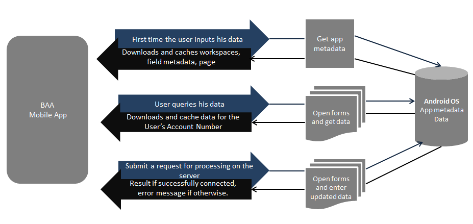
4.3.1 Mobile Application Architecture 29
4.3.2 Used Design Pattern 29
4.4 Application Development 31
4.4.1 Software Pre-requisites 31
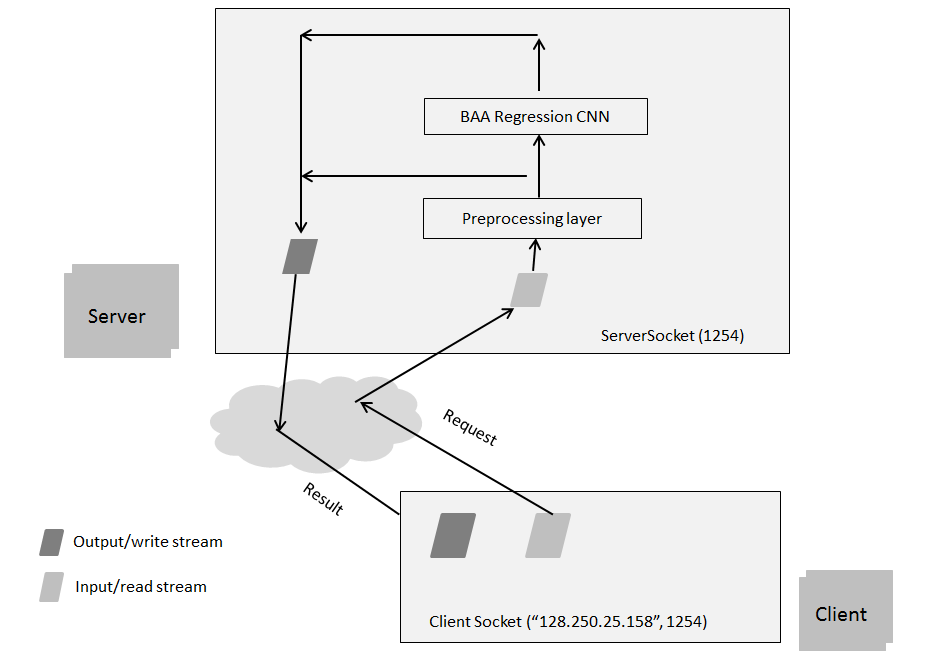
4.4.2 Server 32
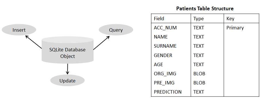
4.4.3 Database 33
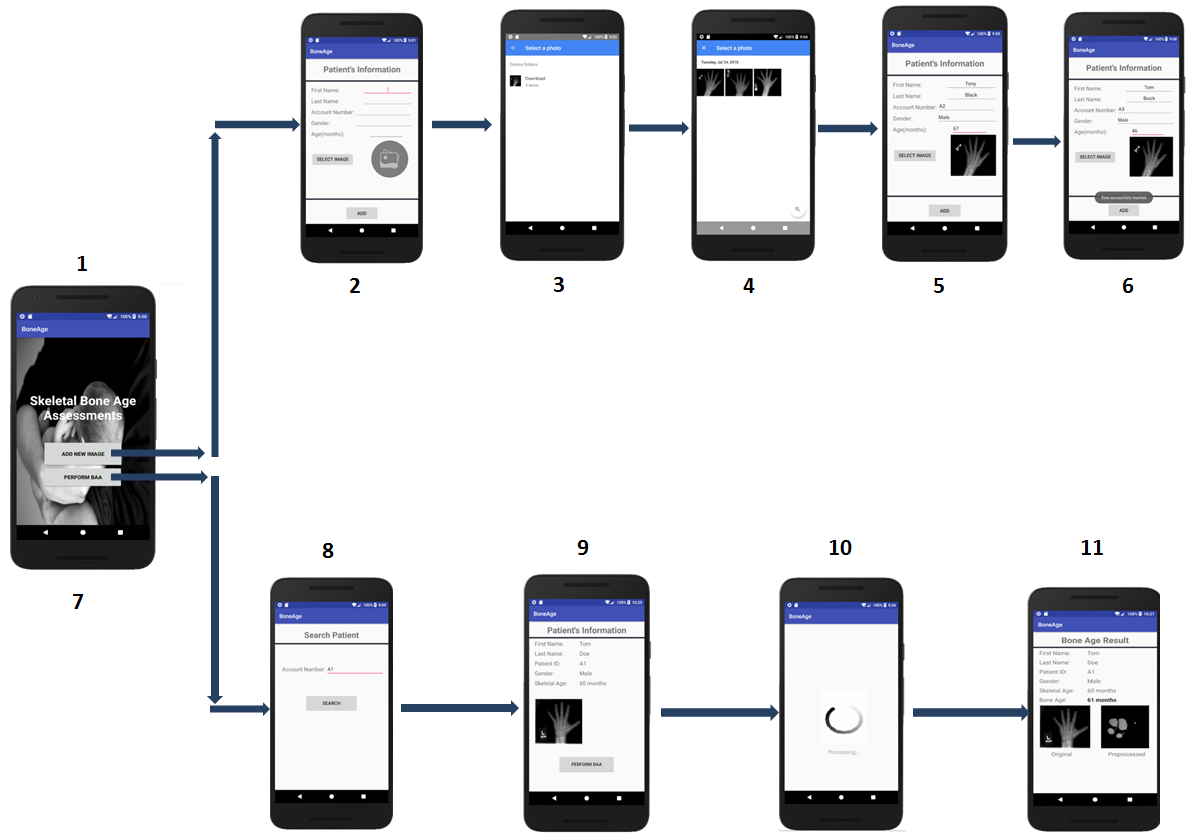
4.4.4 User Interface 34
5. Chapter V BAA Web Application Implementation 38
5.1 Overview 38
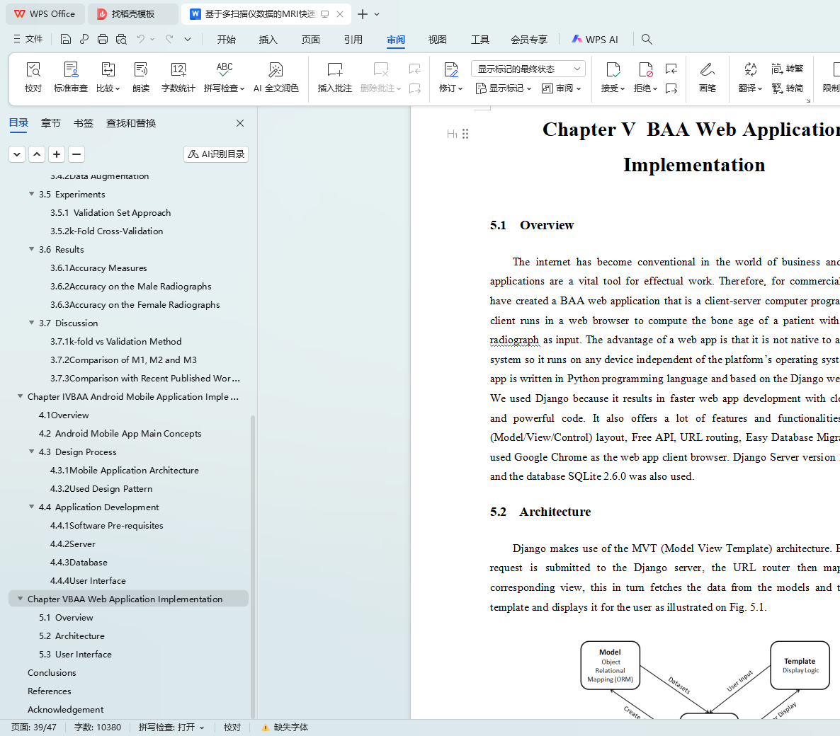
5.2 Web Application Architecture 38
5.3 Web Application User Interface 40
6. Conclusions 42
7. References 43
8. Acknowledgement 47